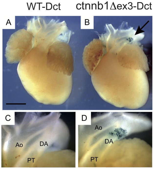
Figure 2. Melanoblasts are numerous in ctnnb1Δex3 DA.

Ventral view of WT-Dct (A) and ctnnb1Δex3-Dct (B) E18.5 hearts stained with X-gal. Note that ctnnb1Δex3-Dct samples contain numerous β-galactosidase-stained cells (arrow) in the ductus arteriosus (DA). High magnification of the WT-Dct (C) and ctnnb1Δex3-Dct (D) DA regions, including the aorta (Ao) and the pulmonary trunk (PT). Scale bar (A, B) = 1 mm.
