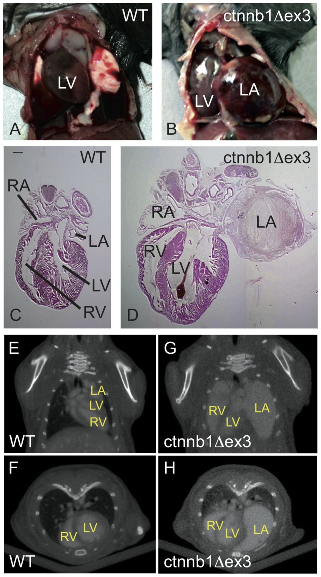
Figure 7. Dilatation of the ctnnb1Δex3 left atrium at postnatal day 28.

Ventral views of Tyr::Cre/°; +/+ ( = WT) (A) and Tyr::Cre/°; ctnnb1Δex3/+ ( = ctnnb1Δex3) (B) open thoracic regions at postnatal day (P)28. Note the size of the ctnnb1Δex3 left atrium. Hematoxylin-eosin staining of WT (C) and ctnnb1Δex3 (D) sections. Note the fibrosis located in the mutant left atrium. Scale bar = 1.5 mm. Frontal (E, G) and transverse (F, H) CT scan pictures of WT (E, F) and ctnnb1Δex3 (G, H) at the truncal level at P28. LA: left atrium; LV: left ventricle; RA: right atrium; RV: right ventricle.
