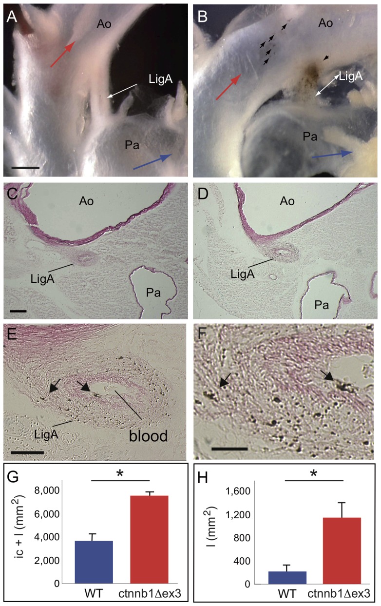
Figure 11. Closure of the ligamentum arteriosum in ctnnb1Δex3 adult heart.

The Tyr::Cre; ctnnb1Δex3/+ ( = ctnnb1Δex3) ligamentum arteriosum (LigA) is not fully closed, rendering it a patent ductus arteriosus. At P28, the LigA does not usually show macroscopic hyperpigmentation in wildtype (WT) mice (arrows, A), whereas Tyr::Cre; ctnnb1Δex3/+ ( = ctnnb1Δex3) LigA does (B). Transverse sections show that the WT LigA (C) is fully closed and does not contain any Mc, whereas ctnnb1Δex3 LigA (D–F) is only partially closed, containing both blood in the lumen and numerous pigmented melanocytes in the wall (E, F). The areas occupied by the intimal cushion (ic) and lumen (l) are shown in G and H, respectively. Cross-sections of the LigA show that the outer tunica is dense, while the inner ellipsoid part, known as the intimal cushion, has a distinct aspect. In ctnnb1Δex3 mice, a lumen is observable inside the ic. Ao: aorta, LigA: ligamentum arteriosum, Pa: pulmonary artery, Mc: melanocyte. Scale bars, (A, B) = 0.25 mm, (C, D) = 100 µm, (E) = 50 µm and (F) = 20 µm. For each genotype, the number of cells were estimated from 8-10 sections per LigA using 4 mice. *: p-value <0.05.
