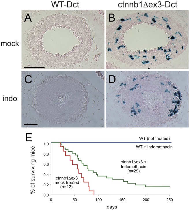
Figure 13. Indomethacin treatment and survival of ctnnb1Δex3 mice.
Indomethacin treatment results in the closure of WT and ctnnb1Δex3 ( = Tyr::Cre/°; ctnnb1Δex3/+) DA and allows the survival of ctnnb1Δex3 mice. Mock (A, B) and indomethacin (indo, 10 mg/kg body weight) (C, D) intraperitoneal injections into pregnant Tyr::Cre/Tyr::Cre; +/+; Dct::LacZ/Dct::LacZ females carrying Tyr::Cre/°; +/+; Dct::LacZ/° (A, C) and Tyr::Cre/°; ctnnb1Δex3/+; Dct::LacZ/° (B, D) E18.5 embryos. Four hours later, embryos were isolated, fixed, X-gal stained, transversally sectioned through the DA and counterstained with eosin. We treated three pregnant females and sectioned ten embryonic hearts (five WT and five mutants). The ductus arteriosus was closed in all cases. Note that the numbers of Dct+ cells derived from ctnnb1Δex3-Dct embryos obtained from pregnant mothers injected with indomethacin or mock-injected were similar. (E) Kaplan-Meier curves of WT and ctnnb1Δex3 newborn pups treated or mock-treated with indomethacin (6 mg/kg body weight indomethacin within 12 hours of birth). Ultrasound analysis was performed on treated versus non-treated animals during the second and third months, which associated survival of treated ctnnb1Δex3 to the size of the left atrium (not shown). Indomethacin-treated ctnnb1Δex3 mice survived significantly longer than mock-treated mice (p<0.009). Note similar results were obtained when ctnnb1Δex3 mi mice were treated with indomethacin or mock. Scale bars, (A, B) = 100 µm, (C, D) = 50 µm.

