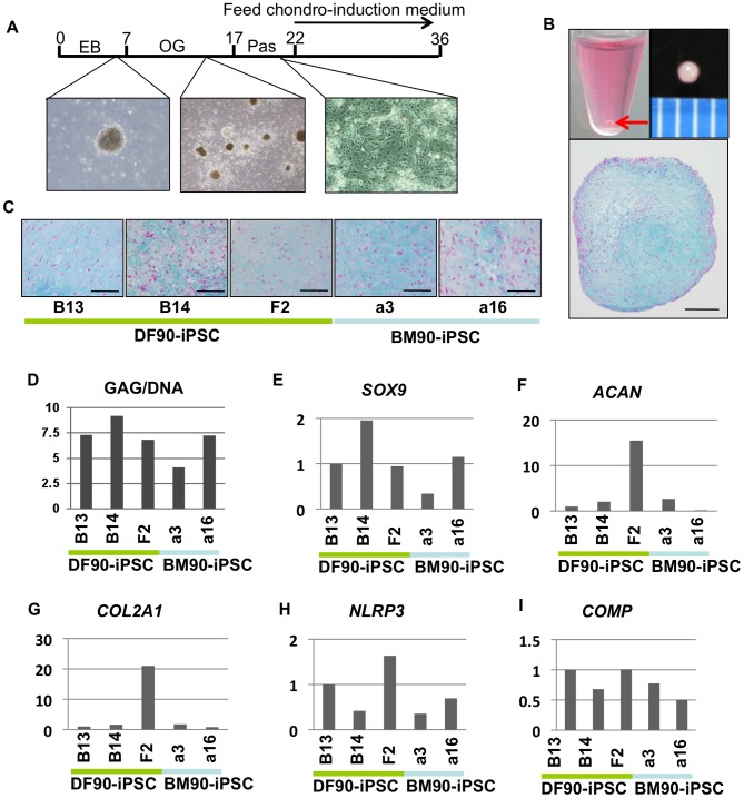
Figure 7. Induction of chondrogenic differentiation in iPSCs.
A) Time course of chondrogenic differentiation. EB, embryoid body. OG, outgrowth. Pas, passaged once. B) Macroscopic views and Alcian blue staining of a section of a pellet. The red arrow indicates a pellet at the bottom of a 15-ml conical tube. Middle column, scale bar, 1 mm. Right column, scale bar, 200 µm. C) Alcian blue staining of sections of pellets. Scale bar, 100 µm. D) GAG/DNA of pellets. GAG/DNA differed with clones regardless of cell-of-origin. E-I) Comparison of the relative expression of chondrogenesis-related genes (SOX9, COL2, ACAN NLRP3 and COMP) by RT-qPCR. The value of DF-iPSC B13 was set to 1 in each experiment.

