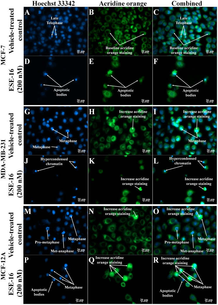
Figure 4. Hoechst 33342 and acridine orange-stained MCF-7, MDA-MB-231 and MCF-12A cells at 400× magnification after 48 h exposure.
Vehicle-treated cells (A–C, G–I and M–O) in various stages of the cell cycle are observed. Formation of apoptotic bodies are observed in ESE-16-treated cells (G–I). An increase in the number of cells in metaphase is observed in ESE-16-treated MCF-12A (P–R) cells when compared to vehicle-treated MCF-12A cells (M–O). An increase in the formation of apoptotic bodies are observed in ESE-16-treated MCF-7 (D–E) and MDA-MB-231 (J–L) cells when compared to vehicle-treated cells (A–C and G–I).

