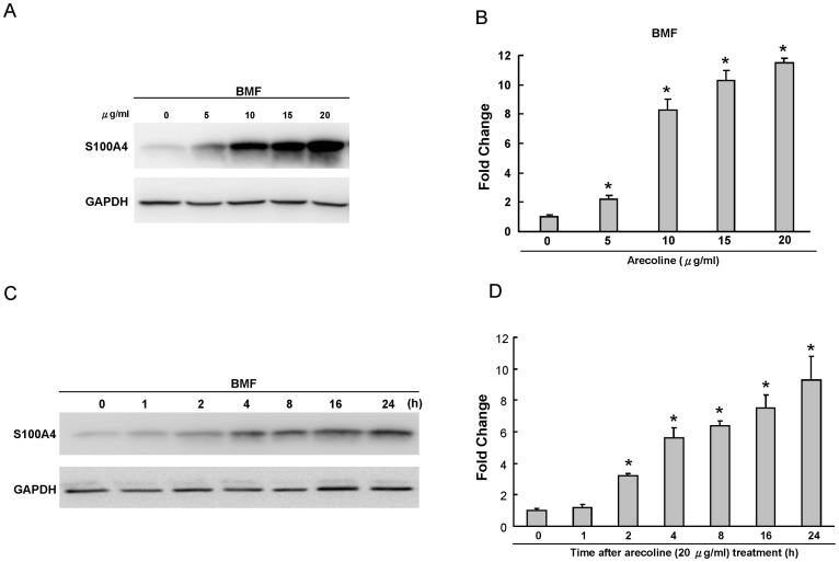
Figure 2. Expression of S100A4 in arecoline-treated BMFs by western blot.
(A) BMFs were exposed for 24 h in medium containing various concentrations of arecoline as indicated. GAPDH was performed in order to monitor equal protein loading. (B) Levels of S100A4 protein stimulated by arecoline were measured by densitometer. The relative level of S100A4 protein expression was normalized against GAPDH signal and the control was set as 1.0. Optical density values represent the mean ± SD. * represents significant difference from control values with p<0.05. (C) Kinetics of S100A4 expression in BMFs exposed to 20 µg/ml arecoline for 0, 1, 2, 4, 8, 16, and 24 h, respectively. GAPDH was performed in order to monitor equal protein loading. (D) Levels of S100A4 protein stimulated by arecoline were measured by densitometer. The relative level of S100A4 protein expression was normalized against GAPDH signal and the control was set as 1.0. Optical density values represent the mean ± SD. * represents significant difference from control values with p<0.05.

