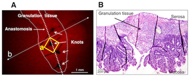
Figure 3. A stereomicroscopic image including the observed site shown in Figure 4 .

A. The thick granulation tissue at the anastomotic region in a mouse that was treated with MOS solution for 1 week after anastomosis surgery. An area in the square (a) corresponds to an area in the square (a) in Figure 4 . B. A microscopic image of a longitudinal section, prepared following fixation, that was taken along the line (b) indicated in panel A.
