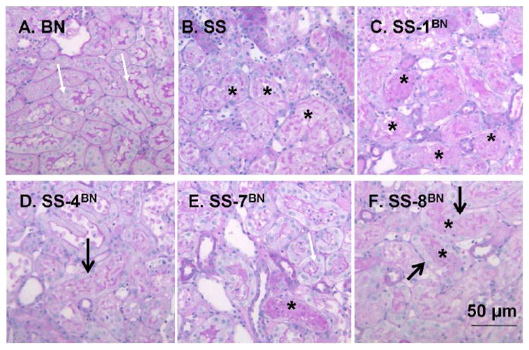
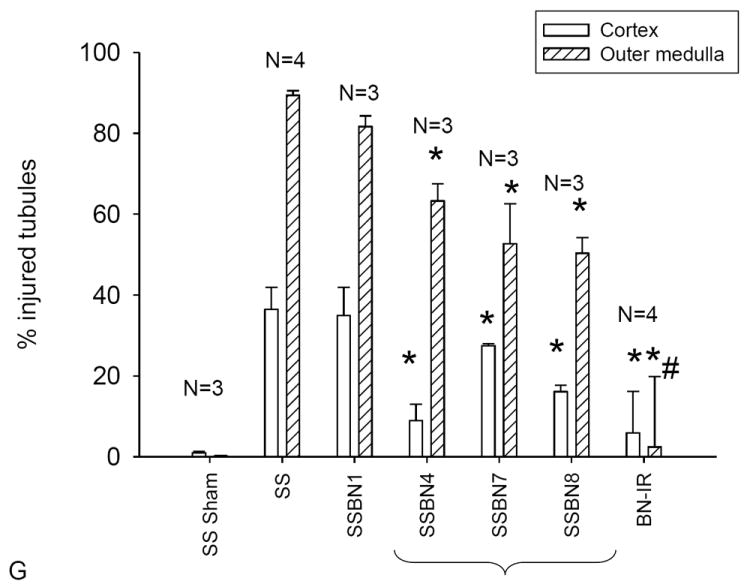
Figure 2.


Representative renal histology of SS, BN and selected consomic rat strains 24 hours following I/R injury. Photomicrographs are shown in the renal outer medulla for the BN (A), SS (B), SS-1BN (C), SS-4BN (D), SS-7BN (E) and SS-8BN (F). White arrows indicate PAS-positive brush border staining, while area of necrotic cellular debris are indicated by *. In some tubules, although cellular debris is present, dedifferentiated and viable cells are clearly seen (thick arrow). Panel G is quantitative analysis of tubular injury scores for renal cortex (open) or outer medulla (hashed bar). Data are expressed as percent affected tubules and are mean ± SE. The N for each group is shown; * P < 0.05 indicates significantly reduced vs SS rats; # indicates injury in BN is less than the consomic protected strains.
