FIGURE 1.
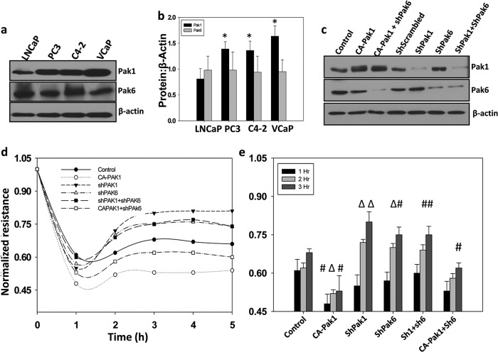
Pak1 controls micrometastasis of prostate cancer cells. a and b, Western blot analysis showing expression levels of Pak1 and Pak6 in human prostate cancer LNCaP, PC3, LNCaP C4-2, and VCaP cell lines normalized to β-actin. c, stable human prostate cancer (PC3) cells expressing shRNA for scrambled (control), Pak1, or Pak6 were made using lentiviral particles following selection with puromycin. Control PC3 cells as well as those expressing ShPak1 and ShPak6 were also transiently transfected with control vector (pBabe-Puro) or constitutively active Pak1 (CA-Pak1, Pak1 E423). c, Western blot analysis of control and transfected PC3 cells with antibodies specific for Pak1, Pak6, and β-actin. d and e, transendothelial migration (microinvasion) of prostate cancer (PC3) cells was measured using ECIS equipment with human dermal microvascular endothelial cells plated on 8W10E+ array chips (14). Control and transfected PC3 cells, detached from the plate by using cell dissociation buffer (20 mm EDTA in PBS (pH 7.4)) to avoid receptor loss because of trypsin digestion, were directly added onto the endothelial cell monolayer at a density of 5 × 10⁴ cells/well in 50 μl serum-free DMEM. Real-time measurements on the transendothelial migration of PC3 cells were recorded by the ECIS up to 5 h. Data are presented as mean ± S.D. n = 3 of triplicate experiments. *, p < 0.001; Δ, p < 0.01; #, p < 0.05 versus control experiments within the same group.

