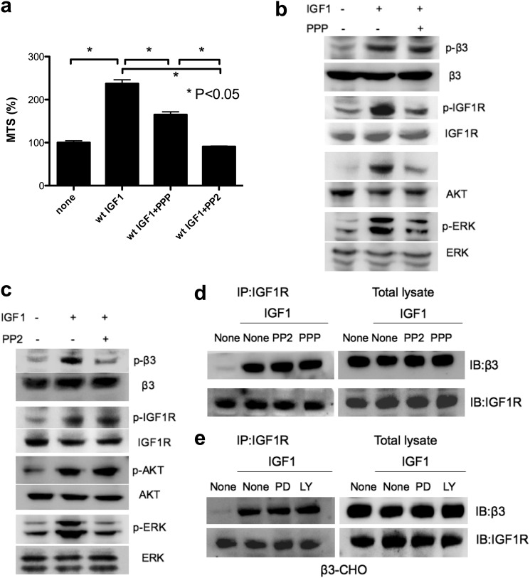
FIGURE 5.
The effect of PPP (IGF1R kinase inhibitor) and PP2 (Src inhibitor) on IGF1 signaling in β3-CHO cells in anchorage-independent conditions. a, the effect of PPP and PP2 on cell survival. β3-CHO cells were plated in polyHEMA-coated plates, serum-starved in DMEM for 48 h, and cultured with IGF1 (100 ng/ml) in the presence or absence of PPP (10 μm) or PP2 (10 μm). Cell survival was measured by MTS assays. The data are shown as mean ± S.E. (n = 6). Statistical analysis was performed using ANOVA and Tukey's multiple comparison test. b and c, the effect of PPP and PP2 on phosphorylation of β3 integrin, IGF1R, AKT, and ERK. p, phospho. β3-CHO cells were plated in polyHEMA-coated plates and serum-starved in DMEM for 3 h. Cells were pretreated with PPP (b) (10 μm) or PP2 (10 μm) (c) for 60 min and then with WT IGF1 (100 ng/ml) for 30 min. Cell lysates were analyzed by Western blotting. d, the effect of PPP and PP2 on ternary complex formation. β3-CHO cells were plated in polyHEMA-coated plates and serum-starved in DMEM for 3 h. Cells were pretreated with PPP (10 μm) or PP2 (10 μm) for 60 min and stimulated with WT IGF1 (100 ng/ml) for 30 min. IGF1R was immunopurified (IP) with anti-IGF1R antibodies from cell lysates, and the immunopurified materials were analyzed with anti-IGF1R or β3 antibodies by Western blotting (IB). e, the effect of PD098059 and LY294002 on ternary complex formation. β3-CHO cells were plated in polyHEMA-coated plates and serum-starved in DMEM for 3 h. Cells were pretreated with PD098059 (50 μm) (PD) or LY294002 (50 μm) (LY) for 60 min and stimulated with WT IGF1 (100 ng/ml) for 30 min. IGF1R was immunopurified with anti-IGF1R antibodies from cell lysates, and the immunopurified materials were analyzed with anti-IGF1R or β3 antibodies by Western blotting.

