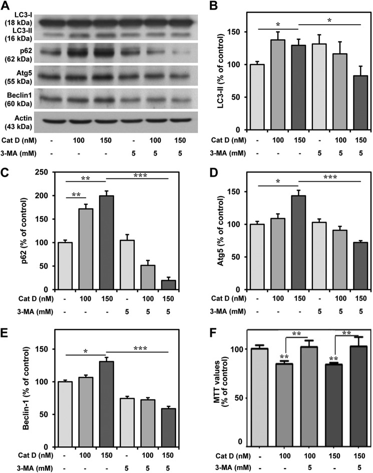
FIGURE 8.
Exogenous cathepsin D increased autophagy markers in cultured neurons. A–E, immunoblots (A) and quantifications (B–E) depicting increased levels of LC3-II, p62, Atg5, and Beclin-1 following treatment with cathepsin D and their reductions with 3-MA treatment in hippocampal cultured neurons. F, MTT assay showing that 3-MA treatment protected cultured hippocampal neurons against cathepsin D-induced toxicity. All results, which are presented as means ± S.E. (error bars), were obtained from three separate experiments. Cat D, cathepsin D; Ctrl, control. *, p < 0.05; **, p < 0.01; ***, p < 0.001.

