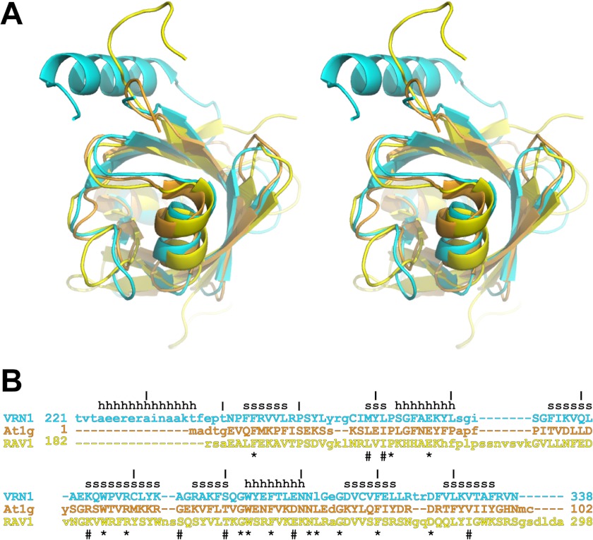
FIGURE 4.
Structural superimposition and structure-based sequence alignment. A, the structures of VRN1(208–341) (cyan), At1g16640 (orange), and RAV1 (yellow) were aligned with SSM in Coot. The resulting structural superimposition is shown in stereo. B, the structure-based sequence alignment of these three proteins (same color scheme as in A) shows structurally aligned residues in uppercase letters. Residues that are conserved in all three structures are shown by asterisks, and residues that are highly conserved are shown by hash symbols. Secondary structure elements in the VRN1(208–341) structure are indicated by h for helix and s for strand. At1g, At1g16640.

