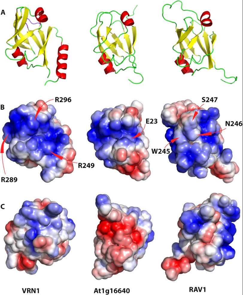
FIGURE 5.
Structural alignment of VRN1, At1g16640, and RAV1 B3 domains. A, structural alignment showing the putative DNA-binding surface of VRN1(208–341) and RAV1. B, electrostatic surfaces of the three domains in the same orientation as in A. Arg residues contributing to the conspicuous basic patch on VRN1(208–341) (R249, R289, and R296) are labeled. C, electrostatic surfaces of the three domains rotated 180° around the vertical axis relative to the surface in B. The structures were first aligned with SSM in Coot and then oriented to show the putative DNA-binding region of RAV1 (9), with the surface charge ranging from −3 to 3 kbT/ec(J/C), where k is the Boltzmann constant (1.380 × 10⁻²³ J/K), T is in Kelvin, and e is the charge on an electron (1.6 × 10⁻¹⁹ C).

