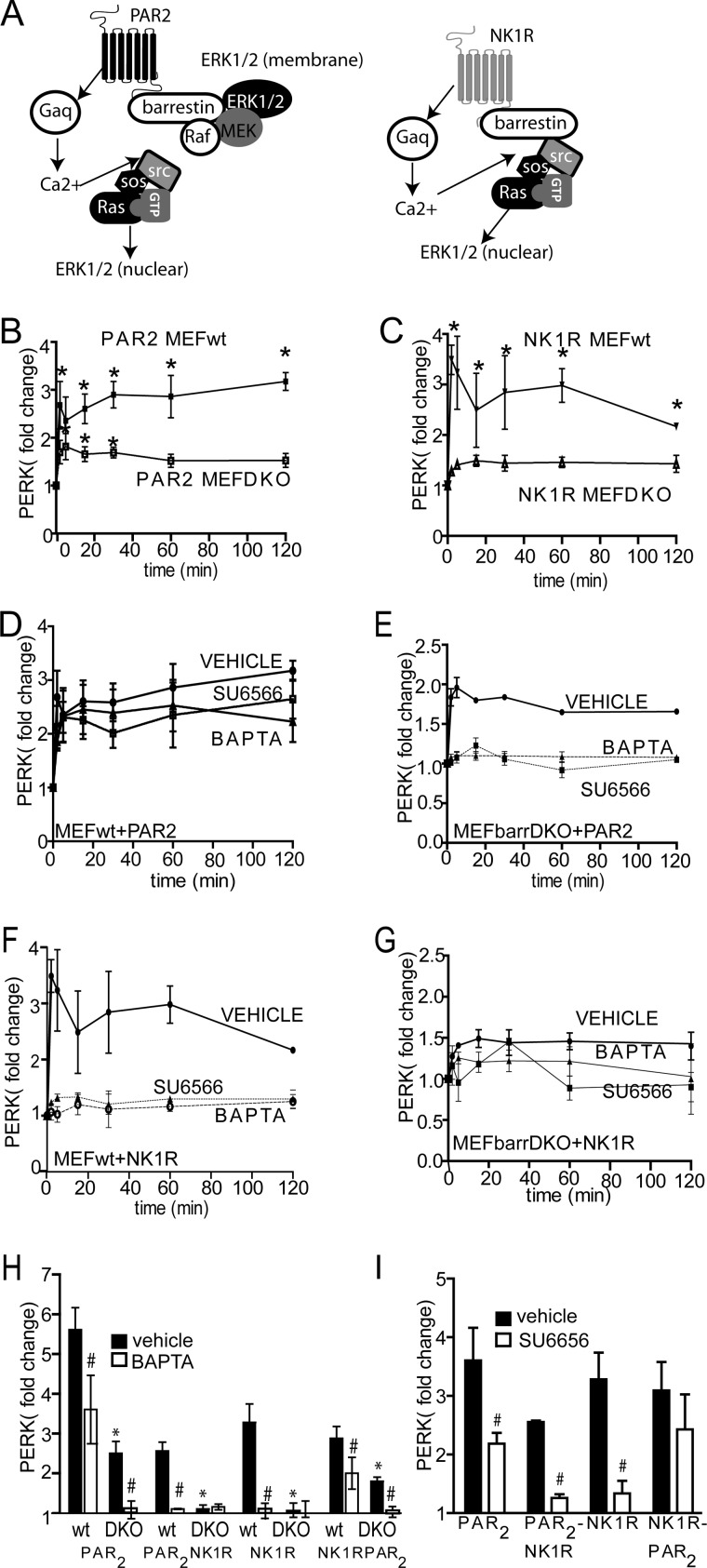
FIGURE 4.
PAR2 and NK1R activate ERK1/2 by distinct β-arrestin-dependent mechanisms. A, schematic depicting signaling to ERK1/2 by PAR2 and NK1R. B and C, mouse embryonic fibroblasts from wild type (MEFwt) or β-arrestin-1/2 knock-out mice (MEFβarrDKO) were transfected with either PAR2 (B) or NK1R (C). Cells were treated with 2fAP or Sar-Met-SP, and ERK1/2 activation was determined by in-cell Western, using pERK and total ERK antibodies. Normalized ERK1/2 activation is graphed as -fold increase over base line (pERK/total ERK in untreated cells). *, statistically significant increase in ERK1/2 phosphorylation compared with base line (p < 0.01, n = 5). ERK1/2 phosphorylation was significantly lower in MEFβarrDKO at all time points (p = 0.001, n = 5). D–G, in-cell Western analysis of ERK1/2 activation in MEFwt (D and F) or MEFβarrDKO (E and G) transfected with either PAR2 (D and E) or NK1R (F and G) after pretreatment with either vehicle (DMSO), BAPTA-AM (to block Ca2+), or SU6566 (Src inhibitor). ERK1/2 phosphorylation was significantly reduced in SU6566- and BAPTA-AM-treated cells compared with vehicle-treated cells at all time points in NK1R-transfected cells and at 5–30 min in PAR2-transfected cells (p < 0.05, n = 5). H, graph of mean maximal agonist-stimulated ERK1/2 activation after pretreatment with vehicle or BAPTA-AM in MEFwt or MEFβarrDKO expressing PAR2, NK1R, PAR2-NK1R, or NK1R-PAR2. I, graph of mean maximal agonist-stimulated ERK1/2 activation in MEFwt, transfected with respective receptor, after pretreatment with SU6566. #, statistically significant difference between vehicle and SU6566- or BAPTA-AM-treated cells. *, statistically significant difference between METwt and MEFβarrDKO (p < 0.05, n = 5). Error bars, S.E.

