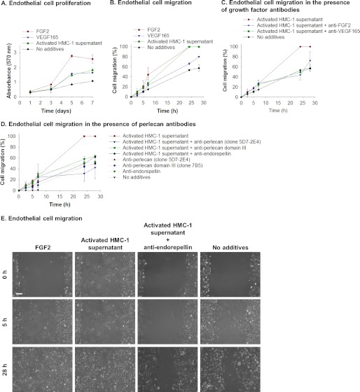
FIGURE 8.
Perlecan promotes endothelial cell migration. A, proliferation of endothelial cells over 7 days in the presence of medium supplemented with FGF2, VEGF165, or activated HMC-1 supernatant compared with cells exposed to medium only. Cell proliferation was measured by crystal violet and presented as mean ± S.D. (error bars) (n = 3). B, endothelial cell migration measured over 28 h in the presence of medium supplemented with FGF2, VEGF165, or activated HMC-1 supernatant compared with cells exposed to medium only. Cell migration was measured by a scratch assay and monitoring the migration of cells into the scratched area. Data are presented as percentage of cell migration, which indicates the scratched area at a given time as a proportion of the initial scratched area. C, endothelial cell migration measured over 28 h in the presence of medium supplemented with activated HMC-1 supernatant, activated HMC-1 supernatant, and anti-FGF2 antibodies (AB1435) or anti-VEGF165 antibodies (ab38473) compared with cells exposed to medium only. Cell migration was measured by a scratch assay and monitoring the migration of cells into the scratched area. Data are presented as percentage of cell migration, which indicates the scratched area at a given time as a proportion of the initial scratched area. D, endothelial cell migration measured over 28 h in the presence of medium supplemented with activated HMC-1 supernatant, activated HMC-1 supernatant, and anti-perlecan antibodies (clone 5D7-2E4 or 7B5 or anti-endorepellin), or anti-perlecan antibodies only (clone 5D7-2E4 or 7B5 or anti-endorepellin) compared with cells exposed to medium only. Cell migration was measured by a scratch assay and monitoring the migration of cells into the scratched area. Data are presented as percentage of cell migration, which indicates the scratched area at a given time as a proportion of the initial scratched area. E, phase-contrast images of the scratched area as times 0, 5, and 28 h after the scratch was generated for endothelial cells exposed to medium supplemented with FGF2, activated HMC-1 supernatant, activated HMC-1 supernatant, and anti-endorepellin compared with cells exposed to medium only. Scale bar, 150 μm.

