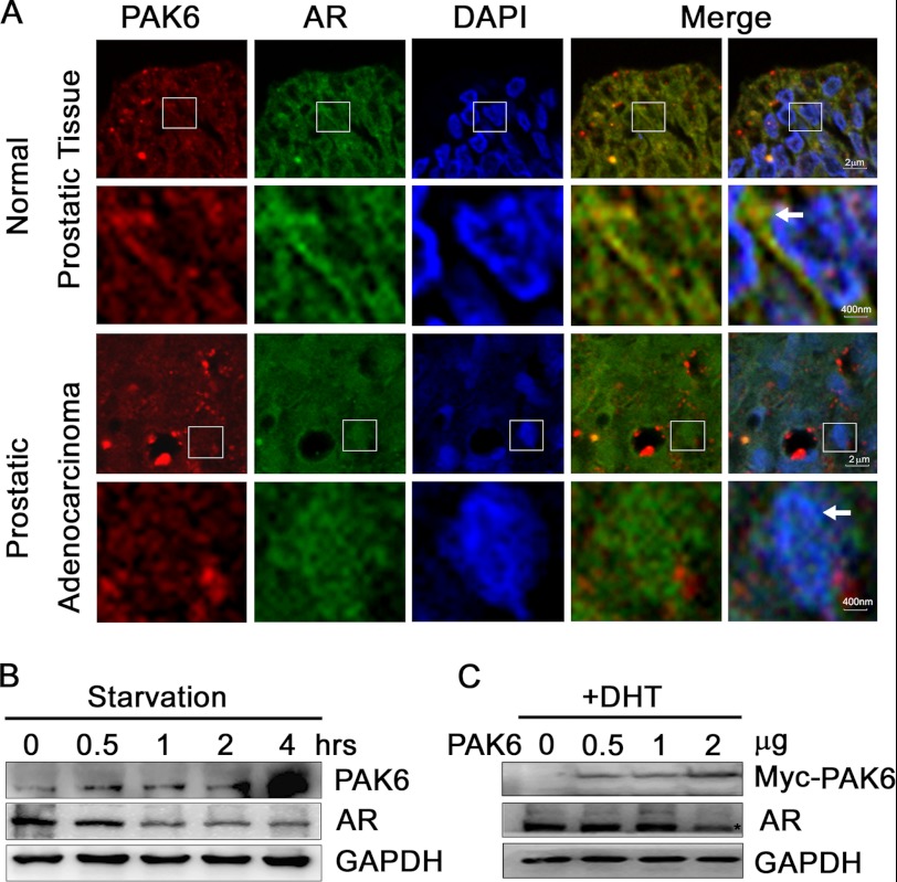
FIGURE 1.
PAK6 involvement in AR localization and expression. A, PAK6 and AR are co-localized in the cytoplasm of normal prostate cells, and AR is localized in the nucleus in prostate cancer cells. Tissue specimens were subjected to immunohistochemical analysis as usual. After antigen retrieval, specimens were fixed and incubated with anti-PAK6 antibody followed by Alexa 594 Fluor (red) antibody and anti-AR antibody followed by Alexa 488 Fluor (green) antibody. Nucleus was stained by DAPI (blue). The 2nd and 4th lines are the 25-fold enlarged pictures of the 1st and 3rd lines, respectively. The 4th row was merged with red and green images, and 5th row was merged with red, green, and blue images. The white arrows in the 2nd line indicate AR co-localizes with PAK6 in the cytoplasm, and arrows in 4th line indicate AR translocates in the nucleus. B, starvation experiment showing that PAK6 expression increased and AR decreased upon starvation. CW22Rv1 cells were starved with Hanks' buffered saline solution (Invitrogen) at different time points as indicated. Western blot analysis was performed, and endogenous proteins were detected with the indicated antibodies. C, CWR22Rv1 cells transfected with Myc-PAK6 show decreased AR levels upon DHT activation. CWR22Rv1 cells were transfected with indicated amounts of pcDNA3.1-Myc-His-PAK6. Endogenous AR and exogenous PAK6 were detected with indicated antibodies.

