FIGURE 3.

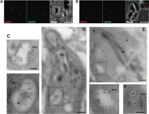
TgCPB resides in the VAC. A and B, newly invaded or overnight replicated RHΔku80 strain parasites were dual-stained with anti-TgCPL and anti-TgCPB catalytic domain antibodies, respectively. TgCPB co-localized with TgCPL in a single VAC of newly invaded parasites (A), whereas it resided in distinct puncta in replicated parasites with a fragmented VAC (B). Bars, 5 μm. TgCPL and TgCPB co-localized in the VAC of an ultrathin sectioned newly invaded parasite (C and D) or replicating parasites (E and F) immunostained with anti-TgCPL (10-nm gold particles) and anti-TgCPB catalytic domain (5-nm gold particles) antibodies. The arrow pinpoints internal membranous structures in the VAC. TgCPB (white circles), and TgCPL (black circle) also showed unique labeling of distinct clusters within the VAC upon fragmentation of this organelle. Bars, 500 nm. Rh, rhoptry.
