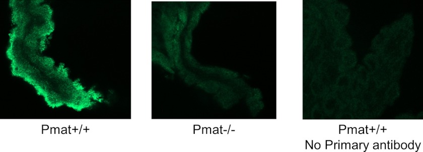
FIGURE 6.
Expression and localization of Pmat in mouse CP. WT and Pmat−/− mouse CPs were isolated, fixed, permeabilized, and immunostained with the P469 anti-PMAT primary antibody and Alexa Fluor 488-conjugated secondary antibody. Confocal scanning sections of immunostained CP tissues are shown. The WT CP tissue without primary antibody incubation was used as a control.

