
FIGURE 1.
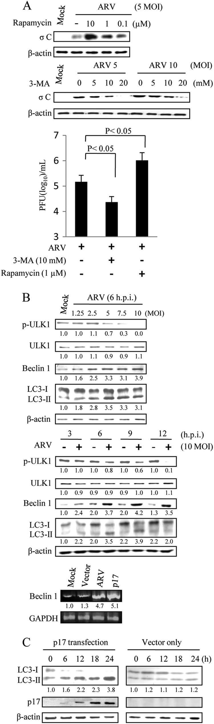
ARV induces autophagy. A, Vero cells were pretreated with different concentrations of 3-MA or rapamycin for 2 h and then infected with ARV at m.o.i. of 10. Cells lysates were separated by SDS-PAGE and immunoblotted with antibodies against ARV σC and β-actin, as indicated. The supernatants of ARV-infected cells in each well were harvested at 24 hpi for viral titration. Each value represents the mean of three independent experiments ± S.D. B, Vero cells were infected with different m.o.i. or time points as indicated. Cells lysates were separated by SDS-PAGE and immunoblotted with antibodies directed against ULK1, LC3-I/II, Beclin 1, and β-actin, as indicated. In semi-quantitative RT-PCR amplification of the Beclin 1 gene, Vero cells were transfected with p17 or infected with ARV at an m.o.i. of 10. In the bottom panel, the p17-transfected or ARV-infected cells were collected at 24 hpi, and total RNAs were extracted for semi-quantitative RT-PCR. After electrophoretic separation in an agarose gel and staining with ethidium bromide, the expression rate of the target gene is assessed by measuring the intensity of the band corresponding to a generated amplicon. The Beclin 1 mRNA levels in ARV-infected and p17-transfected cells were compared with those in mock-treated cells. The mRNA levels were normalized to that for GAPDH. Numbers below each lane are percentages of the control level of a specific protein in the mock treatment. C, Vero cells were transfected with p17-pcDNA3.1 and pCDN3.1, respectively. Cell lysates were harvested at 6, 12, 18, and 24 hpi and immunoblotted with respective antibodies against p17, LC3, and β-actin. Similar results were obtained from three independent experiments.