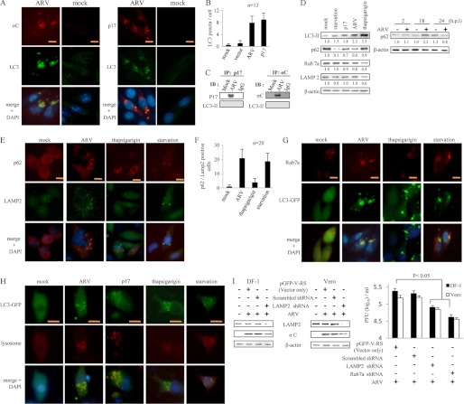
FIGURE 8.
ARV p17 promotes autophagosome and autolysosome formation that benefits virus replication. A, Vero cells were infected with ARV for 12 h and then fixed and processed for immunofluorescence staining of p17, σC, and LC3. The colocalization of ARV proteins (p17 and σC) was observed under a fluorescence microscope. Scale bar, 20 μm. B, quantitation results from Fig. 1 represents mean LC3 puncta per cell. n = 13. C, coimmunoprecipitation of p17, σC, and LC3 was carried out. Cells were infected with ARV at an m.o.i. of 10 and collected 24 h postinfection. About 500 μg of cellular proteins was incubated with 4 μg of anti-p17 or σC antibodies at 4 °C overnight. The immunoprecipitated proteins were separated by SDS-PAGE followed by Western blot, and then proteins were detected with indicated antibodies. Similar results were obtained in three independent experiments. IB, immunoblot. D, uninfected Vero cells were pretreated with TG for 2 h or without TG treatment (mock) in complete media as well as without TG treatment but in amino acid-free media for 2 h (starvation). In parallel experiments, two sets of Vero cells were infected with ARV at m.o.i. of 10 or transfected with p17-pCDNA3.1 plasmid, respectively. Cells lysates were collected at 24 hpi or post-transfection and then analyzed by Western blot with antibodies against p62. E, Vero cells were infected with ARV at m.o.i. of 10 for 12 h and then fixed and processed for immunofluorescence staining of p62 and LAMP2. The procedures for the TG and starvation treatments are described in D. Colocalization of p62 and LAMP2 was observed under a fluorescence microscope. Scale bar, 20 μm. F, p62- and LAMP2-positive cells were quantified by counting the numbers of positive cells in an individual field under a fluorescence microscope. Each value represents the mean of 20 fields ± S.D. G, Vero cells were transfected with LC3-GFP plasmid for 24 h and then infected with ARV at m.o.i. of 10 for 12 h. The cells were then fixed and processed for immunofluorescence staining of LC3-GFP and Rab7a. Colocalization of LC3-GFP and Rab7a was observed under a fluorescence microscope. Scale bar, 20 μm. H, Vero cells were transfected with LC3-GFP plasmid for 24 h and then infected with ARV at m.o.i. of 10 for 12 h. The cells were then fixed and processed for immunofluorescence staining of LC3-GFP and lysosome. In the p17 transfection assay, cells were transfected with p17-pCDNA3.1 plasmid. The treatment conditions for TG and starvation are described as in D. I, Vero cells were transfected with shRNAs targeting LAMP2 or Rab7a as well as scrambled shRNA for 24 h and then infected with ARV at an m.o.i. of 10 for 24 h. Cells lysates were analyzed by Western blot with antibodies against LAMP2, σC, and actin, respectively. The supernatants of LAMP2 and Rab7a knockdown in ARV-infected cells were harvested at 24 hpi for viral titration. Each value represents the mean of three independent experiments ± S.D.

