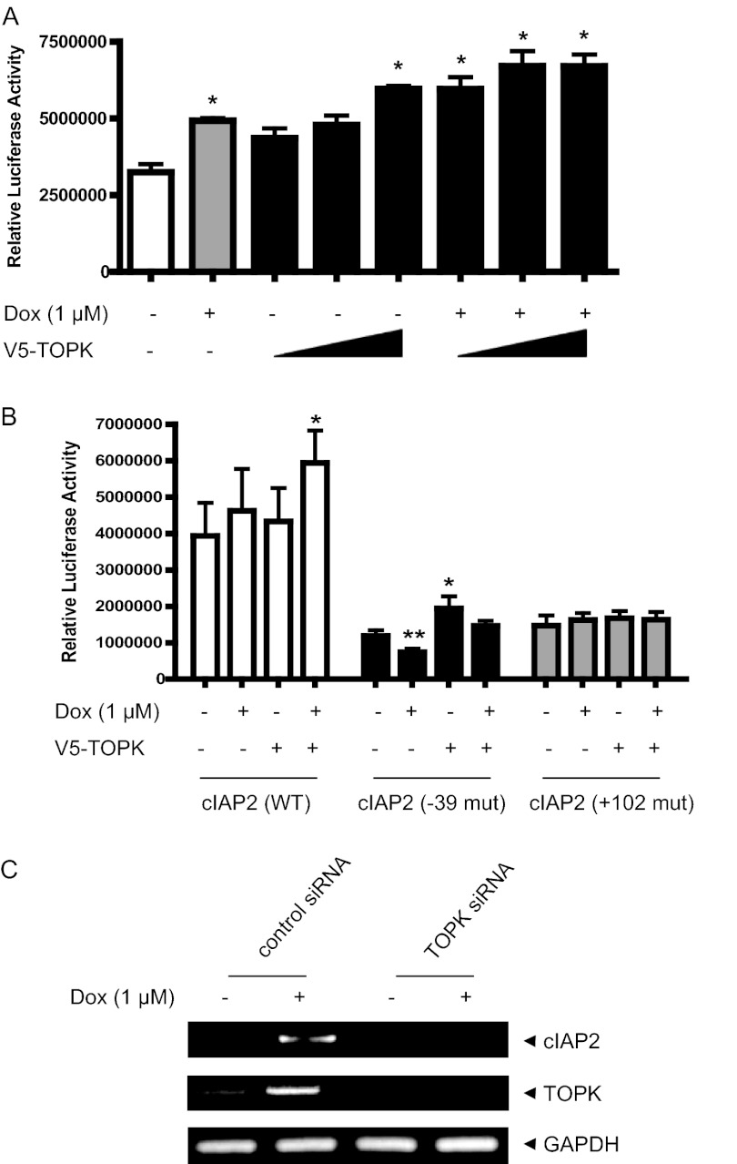
FIGURE 3.
TOPK promotes doxorubicin-induced NF-κB activity and cIAP2 transcriptional activity. A, CHO-K1 cells grown on 6-well plates were transfected with NF-κB-luciferase construct (1 μg) and each indicated V5-TOPK construct (1, 2, 4 μg), together with 0.1 μg of pRL-SV40 gene. 24 h after transfection, the cells were treated or not treated with doxorubicin (Dox,1 μm) for 6 h. Luciferase activity was measured using cell lysate. Firefly luciferase activity was normalized against Renilla luciferase activity. *, p < 0.01; **, p < 0.05. B, CHO-K1 cells grown on 6-well plates were transfected with each WT or mutant (−39 and +102) cIAP2 promoter driven luciferase construct (1 μg) and V5-TOPK construct (1 μg), together with 0.1 μg of pRL-SV40 gene. At 24 h post-transfection, the cells were stimulated with doxorubicin (1 μm) for 6 h. Luciferase activity was evaluated. *, p < 0.01; **, p < 0.05. C, total RNAs were prepared from HeLa-TOPK siRNA or control siRNA cells stimulated with doxorubicin (1 μm). RT-PCR was sequentially performed using SuperScript III reverse transcriptase and each primer for cIAP2 or GAPDH genes. PCR products were resolved on a 1.5% agarose gel and analyzed using GelDoc (Bio-Rad).

