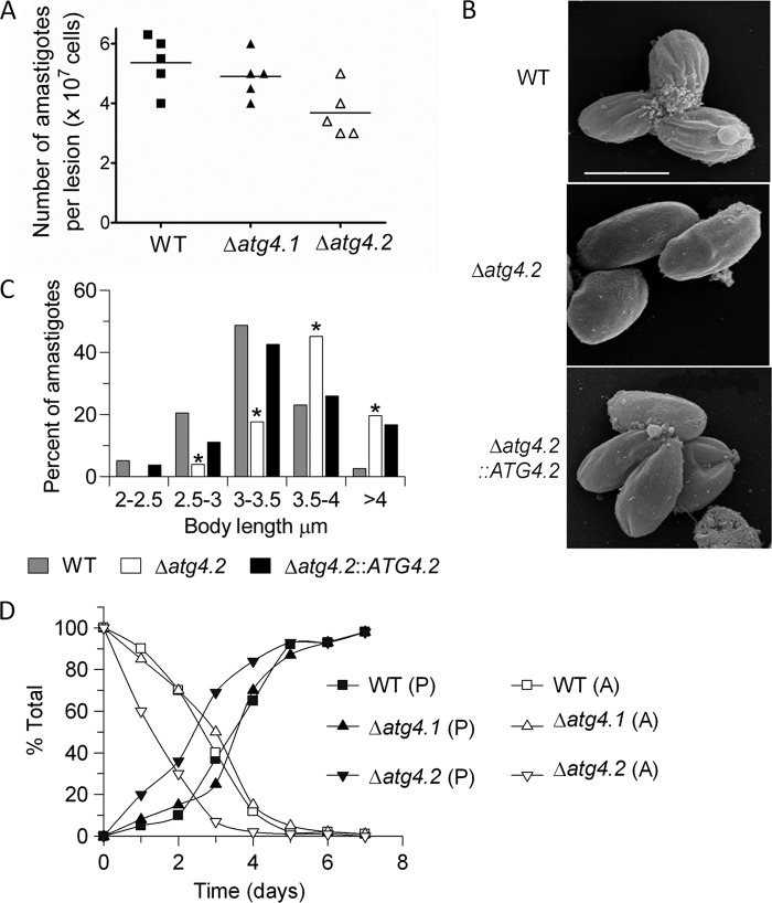
FIGURE 4.
Morphology and transformation of amastigotes of Δatg4.2. A, parasite load in footpad lesions of infected BALB/c mice. The lesions from five mice were dissected when they reached 4 mm in thickness, the tissue was disrupted in PBS, and the number of parasites was determined microscopically. The lines indicate the means, which for WT and Δatg4.1 are significantly different from Δatg4.2, p < 0.05. B, scanning electron micrographs. Bar represents 4 μm. C, histogram of body lengths of amastigotes isolated from lesions in mice. The abscissa shows body length ranges in μm; the ordinate indicates percent of cells counted. *, data for Δatg4.2 differed significantly from those for WT parasites in all class intervals (p < 0.05). D, transformation of amastigotes isolated from BALB/c mice. The data show the disappearance of amastigotes (A) and the appearance of promastigotes (P) over 7 days.

