
Figure 1.
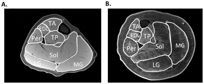
Representative T1-weighted fat suppressed images of the lower leg of a boy with DMD that appears to have relatively little involvement (A) and in a boy that the disease is more progressed (B). This MR sequence can be utilized to determine maximal cross sectional area in various lower leg muscles, including the tibialis anterior (TA), extensor digitorum longus (EDL), tibialis posterior (TP), peroneaus (Per), soleus (Sol), lateral gastrocnemius (LG), and medial gastrocnemius (MG).