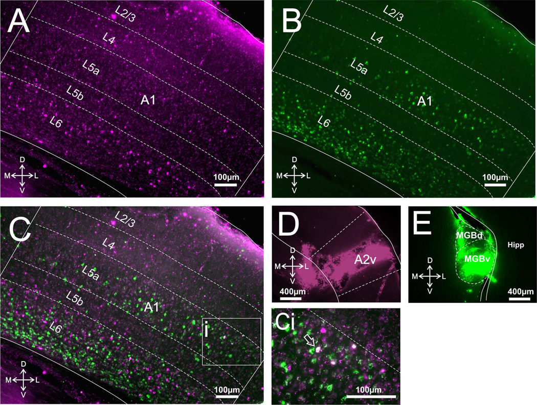
Figure 2.
Corticothalamic and corticocortical cells in A1. A: Photomicrograph of retrogradely labeled cells in A1 following injections of red Retrobeads in A2v. B: Photomicrograph of retrogradely labeled cells in A1 following injections of green Retrobeads in auditory thalamus. C: Overlay of A,B. Ci: Highlighted area in C magnified showing an example of a double-labeled cell in focus, seen in white (arrow). Two more double-labeled cells are also visible but outside the plane of focus. D,E: Photomicrographs of injection sites in A2v and MGB, respectively. MGBv, ventral medial geniculate body; MGBd, dorsal medial geniculate body; Hipp, hippocampus.

