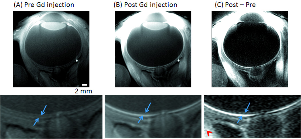
Figure 1.

Anatomical MRI (FLASH) at 0.1×0.2×2 mm from a normal baboon (A) before, (B) after Gd-DTPA administration and (C) the subtracted image. Three distinct “layers” of alternating bright, dark and bright bands are evident. The two (blue) arrows in the expanded views indicate the inner and outer bands of the retina corresponding to the two vascular layers, bounding the retina. The (red) arrowhead indicates signal enhancement of extra-ocular tissues.
