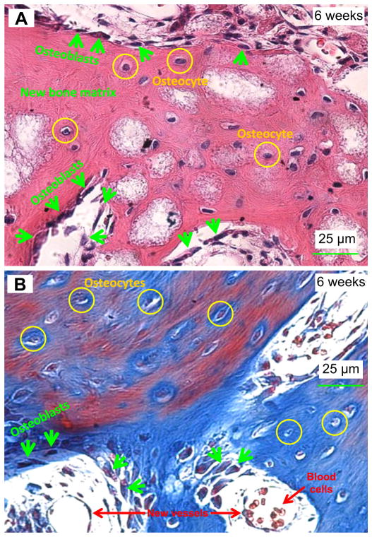
Fig. 4.
Representative H & E (A) and Masson (B) images of tissues retrieved at 6 weeks. Mineralized bone with osteocytes as well as active cubic osteoblasts were revealed. In (A), osteoblasts (arrows) were located at the boundary of the advancing new bone matrix. Osteocytes were embedded in the mineralized bone matrix. In (B), Masson stained bone matrix collagen into blue, and stained mineralization with mature bone into red. Hence, the Masson staining showed a mixture of blue and red regions in (B). New blood vessels and blood cells are indicated in the lower part of (B). (For interpretation of the references to colour in this figure legend, the reader is referred to the web version of this article.)

