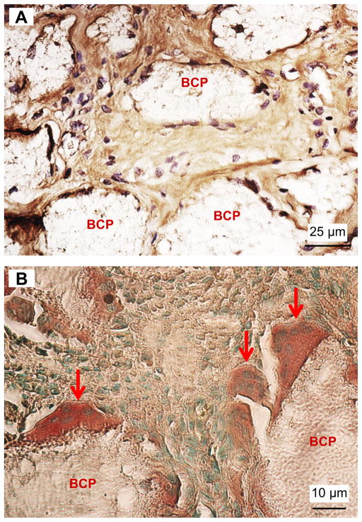
Fig. 5.
Immunohistochemistry staining of collagen I and TRAP activity on 5 μm thick deparaffinized sections of 4 weeks implants. (A) DAB-labeled collagen I (brown) was present inside the BCP implants. (B) TRAP positive multinucleated cells (dark brown) attached to the BCP granules. (For interpretation of the references to colour in this figure legend, the reader is referred to the web version of this article.)

