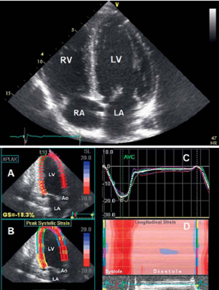
Figure 4.
Above: Echocardiography (four-chamber view) of an athlete’s heart in a male world-class endurance athlete with a heart volume of 19.0 mL/kg body weight. There is a harmonic relationship between the left and right ventricle (LV and RV) and between the left and right atrium (LA and RA).
Below: Echocardiography of an athlete’s heart in a healthy, female, nationally top-ranked endurance athlete with a heart rate of 29 beats per minute at rest.
A: Three-chamber view with wall-motion analysis by 2D speckle tracking.
B: Display of the maximal relative deformation (strain) and shortening of the myocardial segments in systole.
C: The course of relative deformation of the myocardial segments over the cardiac cycle.
D: Display of all analyzed myocardial segments in a three-chamber view (y-axis with color labeling of the segments from B) with synchronous short contraction (red) of all myocardial segments, followed by a long period of relaxation and filling in diastole (pink). LV, left ventricle; LA, left atrium; Ao, aorta

