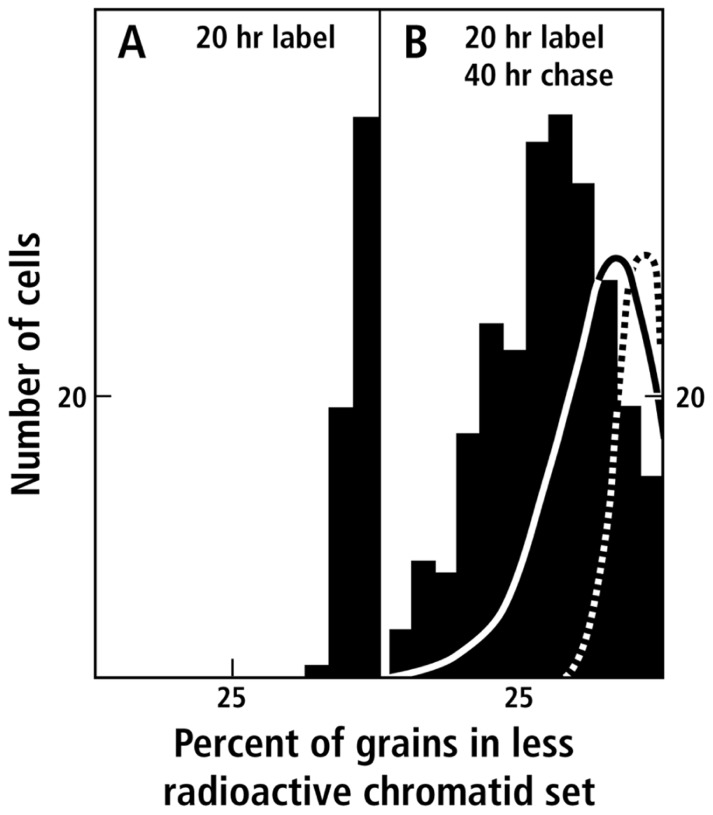
FIGURE 3.
Segregation of radioactively labeled sister chromatids in V. faba root tips (data taken from Figure 3 of Lark, 1967). Radioautographs of anaphase or telophase figures in dividing cells were scored for the number of grains over each sister chromatid set. The distribution of cells is presented as a histogram of the number of cells (ordinate) categorized according to the percent radioactivity in the less radioactive member of the cell’s chromatid pair (abscissa): (A) After 20 h of labeling with radioactive thymidine; (B) after 20 h of labeling with radioactive thymidine followed by 40 h growth in non-radioactive medium. Deviation to the left of 50% indicates the degree of asymmetry. The dashed line indicates the random distribution of 12 radioactive chromatids calculated from the terms of a binomial expansion, assuming no sister chromatid exchange. The solid line is the distribution expected for the random distribution of 12 radioactive chromatids in which sister chromatid exchange results in the redistribution of radioactive material such that on the average 70% of the original chromatid material is not exchanged (for details, see Lark, 1967).

