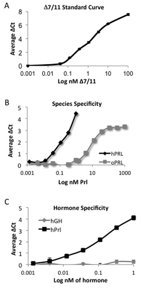
Fig. 2. Optimization of solution-phase proximity ligation assays.

To generate detection probes, SF1a and prolactin receptor antibodies were linked to DNA strands, and upon proximal binding of the antibodies to their respective target proteins, the DNA strands ligate and the PCR amplicon is detected using quantitative real-time PCR. (A) Representative data of Δ7/11 detection using a monoclonal antibody directed against the extracellular domain and the SF1a isoform of the prolactin receptor. (B) Representative data of ovine and human prolactin (oPRL and hPrl, respectively) detection using human prolactin-specific probes. (C) Representative data of growth hormone (hGH) and human prolactin detection using human prolactin-specific probes. Data represent average ΔCt of a minimum of three replicates +/− SD.
