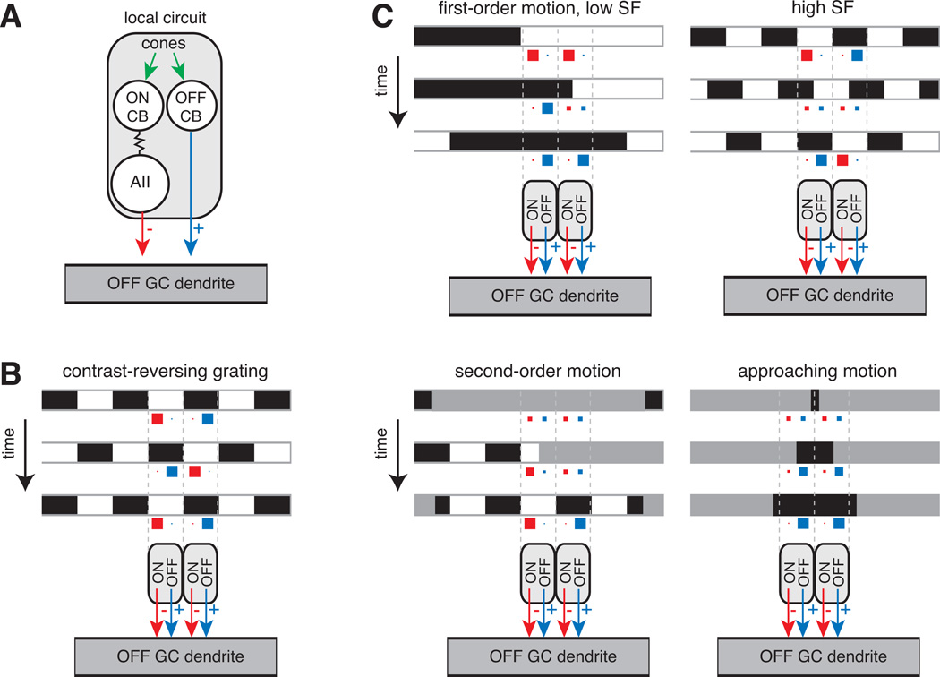
Figure 5. Function of the AII in motion detection.
(A) A local circuit is formed by an OFF cone bipolar cell paired with an ON cone bipolar cell that signals through the AII. Both the OFF bipolar and AII converge on the ganglion cell dendrite. This local circuit is summarized in parts (B) and (C) as an OFF-pathway excitatory input (black arrow) paired with an ON-pathway inhibitory input (red arrow).
(B) Contrast-reversing grating: a high spatial-frequency grating reverses contrast over time. Image frames are shown at three time points. The level of inhibition (red) and excitation (blue) in each of two local circuits is illustrated by the size of squares: larger squares represent higher release rates. On each frame, excitation in one region combines with inhibition in the neighboring region. The summed activity typically results in a burst of firing at each grating reversal showing that excitation dominates (see text).
(C) First-order motion: a drifting grating is shown for both low and high spatial frequency (SF) stimuli. Bars move rightward and stimulate both OFF bipolar and AII amacrine cell release. At the low spatial frequency, excitation and inhibition alternate; whereas at the high spatial frequency, excitation and inhibition co-occur. In different experiments (see text), the high spatial frequency stimulus generates either a net excitation or a cancellation at the ganglion cell level.
Second-order motion: a stationary grating’s contrast is modulated by a moving contrast boundary. The OFF bipolar and AII cell are stimulated as the low contrast boundary moves rightward, revealing the underlying stationary grating. The combination of these inputs generates a modulation of membrane potential around rest and drives firing (see text).
Approaching motion: a small dark stimulus expands over time. The stimulus drives increased release from OFF cone bipolars while suppressing release from AIIs leading to high firing rates in the ganglion cell (see text).

