Fig. 5.

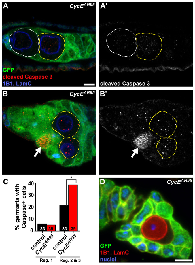
CycE mutant GSCs are not lost through apoptosis. (A-B′) Mosaic germaria showing GFP-negative CycEAR95 clones. GFP (green), wild-type control cells; cleaved Caspase 3 (red) (greyscale images in A′,B′), early apoptosis marker; 1B1 (blue), fusomes; LamC (blue), cap cell nuclear envelopes. CycEAR95 GSCs are outlined in white; CycEAR95 cystoblast-like cells are outlined in yellow. Arrows indicate a GFP-positive wild-type germ cell positive for cleaved Caspase 3. (C) Percentage of germline-mosaic germaria with at least one Caspase-positive cell in region 1 (which contains GSCs and cystoblasts) or regions 2 and 3 (which normally contain more differentiated cysts) in ‘mock’ control (black) or CycEAR95 (red) mosaic germaria. Numbers in bars represent number of germline-mosaic germaria analyzed. *P<0.01. (D) Single GFP-negative CycEAR95 germ cell enveloped by follicle cells in mosaic ovariole. GFP (green), wild-type control cells; 1B1 (red), follicle cell membranes. LamC (red) stains germ cell nuclear envelope; DAPI (blue), nuclei. Scale bars: 5 μm.
