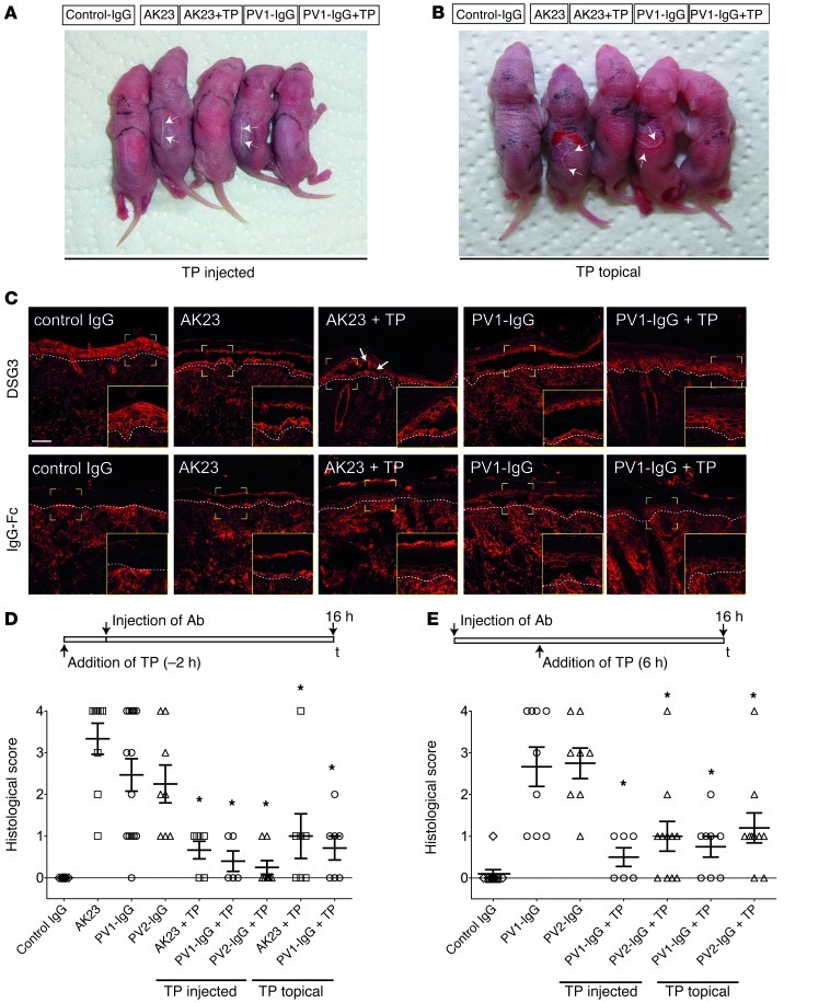
Figure 1. TP blocked acantholysis in vivo.
TP blocked macroscopic blistering (arrows) when injected into the back skin of neonatal mice (A) or applied topically (B). Processing of cryosections revealed typical suprabasal blistering under conditions of AK23 and PV1-IgG injections, but absence or minor blistering (arrows) under conditions of topical TP treatment (C, upper panels). Lower panels demonstrate proper IgG deposition within the epidermis following injection of AK23 or PV-IgG and absence of staining in control-IgG–injected animals. Dashed lines represent dermal-epidermal junction. Scale bar: 50 μm (inserts, ×2 magnification). Evaluation of blister size in serial sections under conditions of TP application 2 hours prior to Ab injection (D) and 6 hours after Ab injection (E). Serial sections of skin samples were evaluated for cleft length and sorted into a score ranging from 0 to 4 as detailed in Methods. Every data point represents 1 injected animal; higher values indicate stronger blistering. *P < 0.05 Ab injection vs. respective Ab injection + TP.

