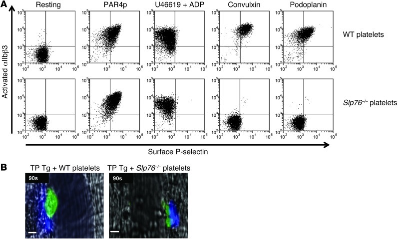
Figure 5. Impaired platelet ITAM signaling and thrombosis in Slp76–/– mice.
(A) Washed platelets from WT or Slp76–/– mice were activated with the indicated agonists, stained for activated αIIbβ3 and surface P-selectin, and analyzed by flow cytometry. Data are representative of 3 independent experiments. (B) Representative images of cremasteric venules taken 90 seconds after laser injury in TP hIL-4Rα/GPIbα–Tg mice reconstituted with 8 × 10⁸ WT or Slp76–/– platelets. Mice were injected with Alexa Fluor 488–labeled antibodies against GPIX to label circulating platelets (green) and Alexa Fluor 647–labeled antibodies against fibrin (blue). Scale bars: 10 μm.

