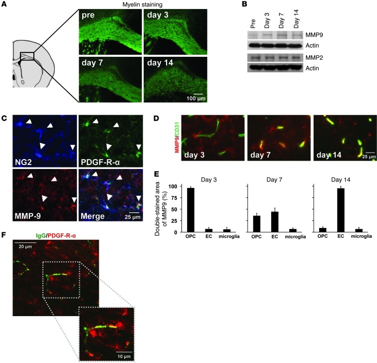
Figure 1. OPCs and MMP9 under white matter pathology.
(A) Cerebral prolonged hypoperfusion stress–induced demyelination in the mouse corpus callosum. n = 5. Quantitative data are shown in Supplemental Figure 1. (B) In our white matter injury model, MMP9 but not MMP2 was increased in the white matter. (C–E) At day 3, most MMP9 signals were observed in NG2/PDGF-R-α–positive OPCs. But at later time points, at days 7 and 14, CD31-positive cerebral endothelial cells (EC) were colocalized with MMP9 signals. n = 5. (F) Notably, OPCs (PDGF-R-α) existed around BBB leakage areas (IgG) at day 3.

