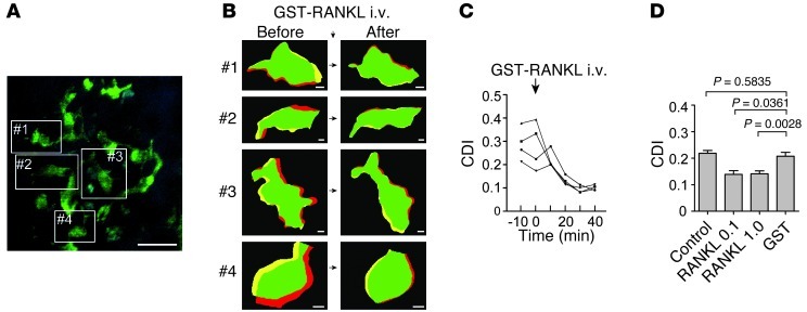
Figure 4. RANKL-mediated rapid control of mature osteoclast function.
(A) Intravital multiphoton imaging of osteoclasts in mouse bone tissues of a3-GFP mice under control conditions (Supplemental Video 6). Mature osteoclasts expressing GFP-fused V-type H+ ATPase a3 subunit are in green. Blue, bone surface. Cell borders are marked in white lines. Scale bar: 40 μm. (B) Representative computer-processed images of mature osteoclasts and their RANKL-mediated rapid shape changes. Images 1–4 were computer extracted from images under the initial condition from A (left panels) and again 10 minutes after i.v. injection of 1 mg/kg of GST-RANKL (right panels) (as in Figure 1, E and F). Scale bars: 5 μm. (C) Representative time courses of the CDI for the 4 individual cells shown in B. Moving (high CDI) osteoclasts underwent transition to the static (low CDI) state less than 10 minutes after i.v. administration of RANKL. (D) The summary of CDIs under control conditions (control) and 40 minutes after i.v. injection of 0.1 mg/kg of GST-RANKL (RANKL 0.1), 1 mg/kg of GST-RANKL (RANKL 1), or GST alone (n = 13 for control, n = 5 for 0.1 mg/kg of GST-RANKL, n = 13 for 1 mg/kg of GST-RANKL, and n = 21 for GST alone, compiled from 3 independent experiments).

