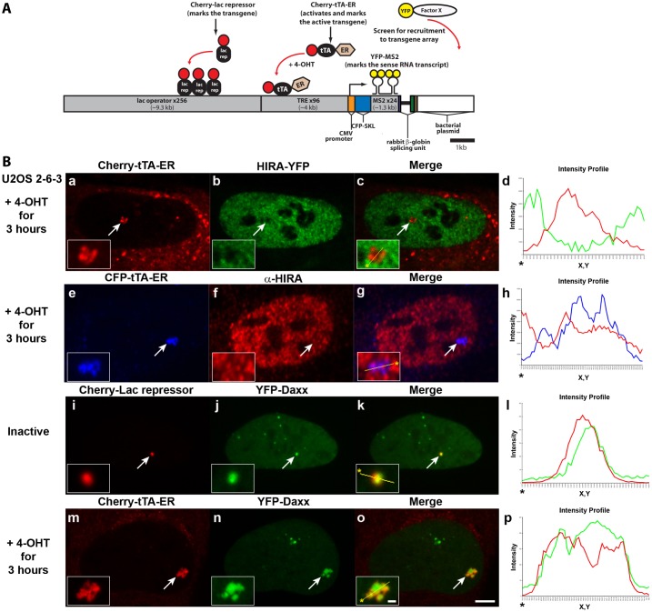
Fig. 1.
The histone H3.3 chaperone, Daxx, is recruited to the transgene array in U2OS cells. (A) Diagram of the inducible transgene drawn to scale. Expression of Cherry-lac repressor allows the inactive transgene array to be visualized. Transcription is induced by the activator, Cherry-tTA-ER, in the presence of 4-hydroxytamoxifen (4-OHT). The transcribed RNA encodes CFP fused to a peroxisomal targeting signal (SKL). The RNA is visualized by YFP-MS2, which binds to the stem loops in the transcript. The 3′ end of the transcription unit is the intron 2 splicing unit of the rabbit β-globin gene. The recruitment of YFP-tagged factors to the array can be monitored by co-expression with Cherry-lac repressor or Cherry-tTA-ER. (B) Localization of HIRA-YFP (a–d) and endogenous HIRA labeled with α-HIRA antibody (e–h) in relation to Cherry- or CFP-tTA-ER, at the activated transgene array in U2OS 2-6-3 cells. YFP-Daxx is enriched at the inactive array, marked by Cherry-lac repressor (i–l), and the activated array, marked by Cherry-tTA-ER (m–p). Yellow lines in enlarged merge insets show the path through which the red, green and blue intensities were measured in the intensity profiles (d, h, l and p). Asterisks mark the start of the line. Scale bar: 5 µm; inset: 1 µm.

