Fig. 2.
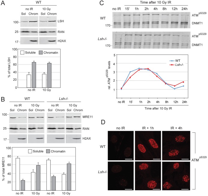
Normal activation of DNA damage response in wild-type (WT) and Lsh−/− MEFs. (A) Western blot detecting LSH before and after IR in the soluble (Sol) and chromatin-bound (Chrom) fractions of nuclear proteins. Quantification of western blots from several independent experiments indicates that before and after IR, ∼60% of LSH is constitutively bound to DNA/chromatin and ∼40% is in the soluble nucleoplasm fraction. RAN and H2AX serve as loading controls for the soluble and chromatin fractions, respectively. (B) MRE11 accumulates equally well on chromatin after IR in wild-type and Lsh−/− MEFs and is partly depleted from the soluble nucleoplasm fraction. The bar graph shows quantification of MRE11 in the soluble and chromatin fractions from three independent experiments. The error bars in A and B represent S.D. (C) The activating phosphorylation of ATM at Serine 329 (pS329) displays similar kinetics after irradiation of the wild-type and Lsh−/− MEFs. DNMT1 is a loading control. The numbers on the left indicate molecular weight in kDa. The graph shows quantification of phosphorylated ATM relative to DNMT1 during the time course. (D) Phosphorylated ATM is recruited to DNA damage foci after irradiation of the wild-type and Lsh−/− MEFs. The scale bars represent 10 µm.

