Fig. 5.
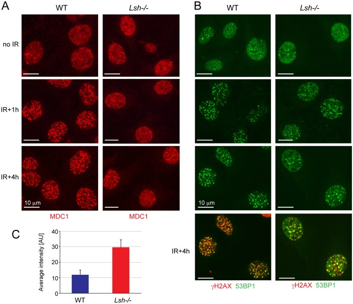
Impaired recruitment of MDC1 and 53BP1 to the sites of DNA damage in Lsh−/− MEFs. (A) Immunostaining of non-irradiated and irradiated wild-type (WT) and Lsh−/− MEFs with antibodies detecting γH2AX binding protein MDC1. Note that diffuse staining for MDC1 can be seen in Lsh−/− MEFs 1 and 4 hours after exposure to IR. (B) Immunostaining of non-irradiated and irradiated wild-type and Lsh−/− MEFs with antibodies against 53BP1. Similar to MDC1, there is more diffuse staining for 53BP1 in Lsh−/− MEFs at 1- and 4-hour time points. The co-localisation of 53BP1 with γH2AX shown in the bottom two panels clearly shows diffuse 53BP1 staining in Lsh−/− MEFs that does not overlap with γH2AX. Note the reduced number of γH2AX foci in Lsh−/− MEFs at this time point in comparison to the wild-type MEFs. The scale bars represent 10 µm. (C) Quantification of 53BP1 staining in the nucleoplasm in wild-type and Lsh−/− MEFs 4 hours post-IR. The error bars represent S.D.

