Fig. 6.
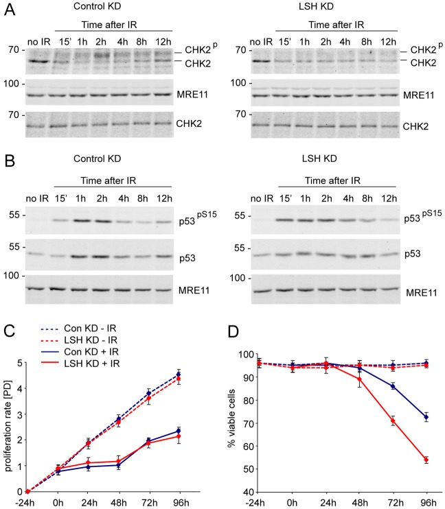
Reduced phosphorylation of CHK2 in LSH-deficient cells. (A) Western blots detecting CHK2, phosphorylated CHK2 and MRE11 in nuclear extracts of non-irradiated and irradiated control KD and LSH KD MRC5 cells. Note that the slower migrating, phosphorylated form of CHK2 is barely detectable throughout the time course of recovery after IR in the LSH KD cells. Extracts prepared without phosphatase inhibitors (bottom panels) do not show the slower migrating phosphorylated form of CHK2. (B) The control KD and LSH-deficient cells display stabilisation of p53 and ATM-dependent phosphorylation of p53 at Serine 15 (pS15) after IR. MRE11 serves as a loading control. The numbers on the left indicate molecular weight markers in kDa. (C) Proliferation of non-irradiated (−IR) and 2.5 Gy irradiated (+IR) control and LSH KD MRC5 cells at indicated time points. ‘[PD]’ indicates population doublings. (D) Viability of non-irradiated and irradiated control and LSH KD cells as detected by trypan blue staining at indicated time points. The error bars represent standard error of the mean.

