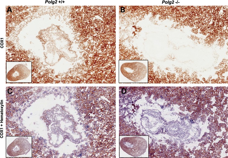
Figure 4.
COXI staining was used to determine mitochondrial function. Ten micrometer cryostat sections of embryos at E8.5 post coitis still enveloped in the maternally derived, deciduum at ×10 magnification and the inset panel at ×2.5 magnification. (A) A Polg2+/+ embryo and the maternal deciduum both stained positive for COXI activity. (B) A Polg2−/− embryo lacking COXI activity. (C) A Polg2+/+ embryo counterstained with hematoxylin showing a dual staining with COXI-positive cells. (D) A Polg2−/− embryo counter stained with hematoxylin showing COXI-negative cells stained in purple revealing the presence of an embryo in the deciduum.

