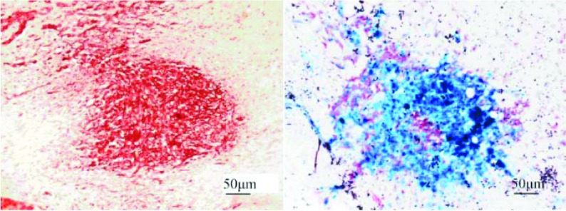
Figure 2. Photomicrographs of LacZ-labelled MSCs injected intravenously after transplantation.
The sections were co-stained with X-Gal (5-bromo-4-chloroindol-3-yl β-D-galactopyranoside) (allowing the LacZ-expressing MSCs to stain blue) and with Neutral Red (allowing the elongated glioblastoma cells to stain dark red. X-Gal staining showed a large number of LacZ-labelled MSCs aggregated in the tumour bed (right-hand panel, ×40). The blue MSCs infiltrated into the bed and scattered throughout the tumour 7 days after injection.

