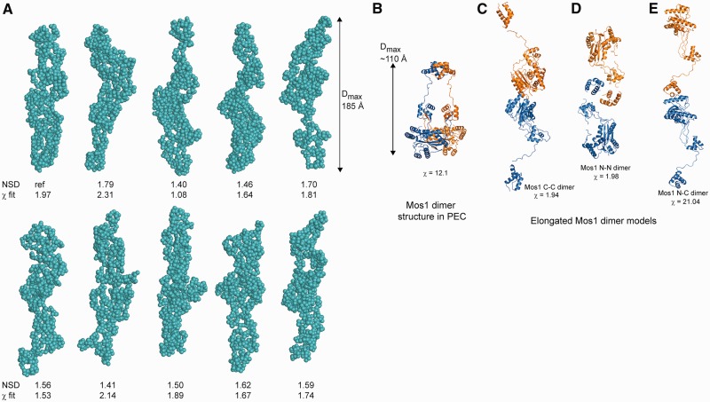
Figure 3.
Solution conformations of Mos1 transposase. (A) Gallery of ten spherical bead models of the H-Mos1 dimer calculated from the SAXS data in GASBOR, with no symmetry imposed. The χ of the fit to the experimental SAXS data and the NSD between models is indicated below each model. (B) Compact structure of the Mos1 dimer in the PEC crystal structure (from PDB ID: 3HOT); one Mos1 monomer is blue and the other orange. (C) An elongated tail-to-tail Mos1 dimer with a catalytic domain dimerization interface (C-C model). (D) Alternative elongated head-to-head model with a DNA-binding domain interface (N-N model). (E) The elongated head-to-tail (N-C) model fitted poorly to the scattering data.

