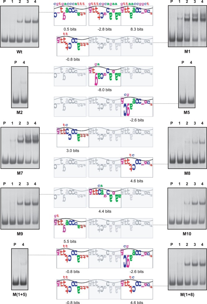
Figure 6.
Sequence walker analysis of the PhoP-binding sites in the non-coding strand of glnA promoters. Sequence walkers (35) serve, for individual sequences, to show the contribution of each base to the conservation of the DRu. The full-length sequence walker (top) corresponds to the wild-type promoter; only the modified DRus are shown for the mutants, with the changed nucleotides over the walker. Analyses by EMSA of the promoters are shown in both sides. P, probe without protein; 1–4, increasing concentrations of GST-PhoPDBD protein (from 0.125 µM to 1 µM). For M2, M5 and M(1 + 5) promoters only the highest concentration (1 µM) is shown.

