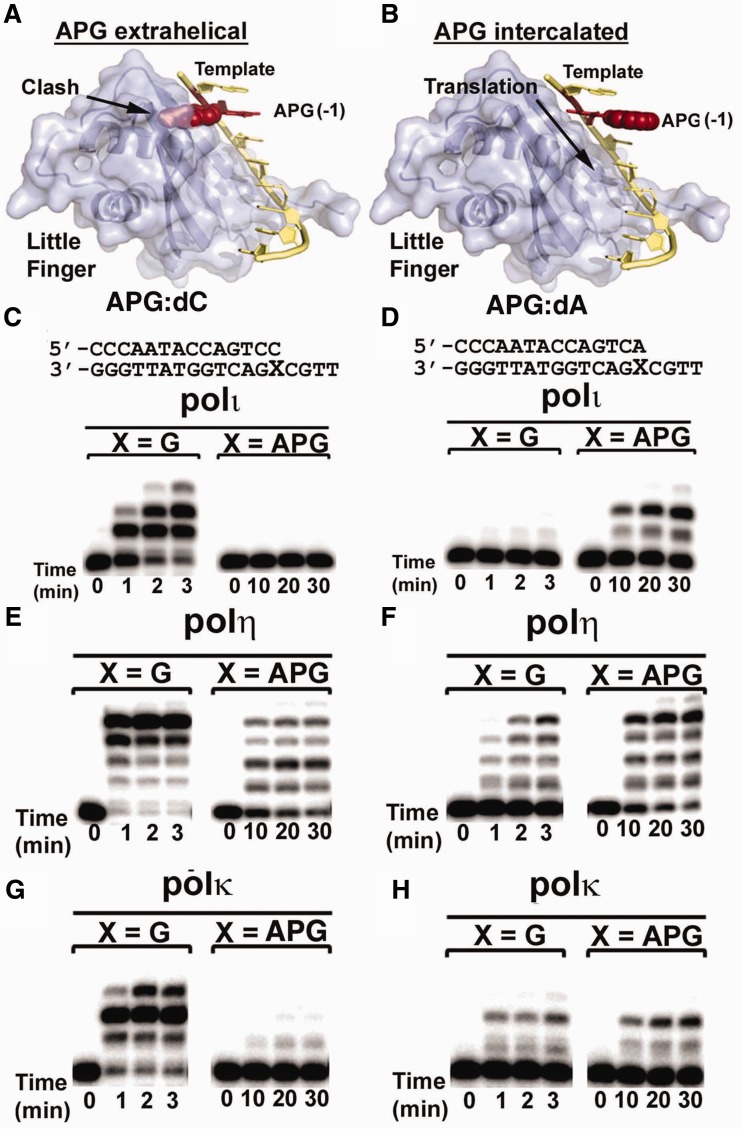
Figure 5.
Effects of APG conformations on primer extension beyond APG lesion. Modeling of template DNA translocation through polι with extra-helical (A) and intra-helical (B) APG conformations. Template DNA is shown in yellow, the APG lesion at the -1 extension position is shown in red and the little finger domain is shown in light blue. Polι, polη and polκ were incubated with undamaged G or the APG lesion paired with correct C (C, E, G) or mismatched A (D, F, H) from the primer strand. Reactions were carried out with all four nucleotides at various time points, as indicated under each lane. DNA substrates are shown above the gels.

