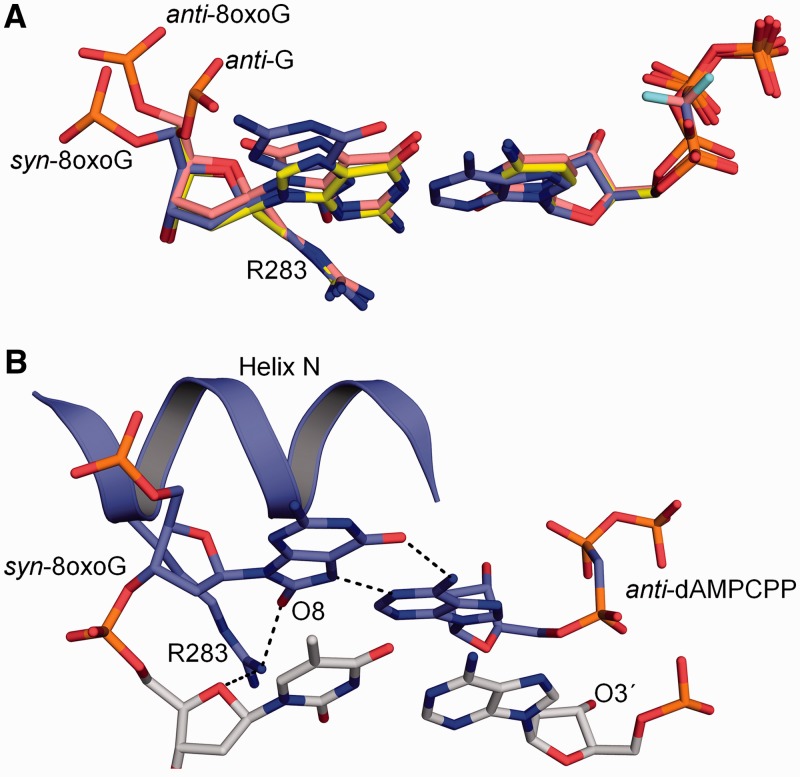
Figure 1.
Structural overview of wild-type pol β with templating 8-oxoG in the active site. (A) Overlay of the templating anti-G:dCMPCPP (PDB ID 2FMP), anti-8-oxoG:dCMP(CF2)PP (PDB ID 3RJI) and syn-8-oxoG:dAMPCPP (PDB ID 3RJF) in the pol β active site are shown in yellow, salmon and purple, respectively. The position of the templating phosphate backbone is indicated for each conformation. (B) Structure of the closed ternary pol β complex with a templating syn-8-oxoG Hoogsteen base pairing with dAMPCPP (PDB ID 3RJF) (7). The primer terminus (O3′) is indicated. Key hydrogen bonding interactions are shown as black dashes.

