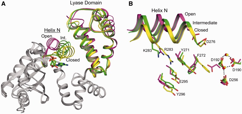
Figure 5.
Intermediate conformation of the R283K pol β ternary complex with templating anti-8-oxoG and incoming dAMPCPP. (A) Structural overlay of the R283K anti-8-oxoG:dAMPCPP ternary complex with the wild-type open binary (PDB ID 3ISB) and closed ternary (PDB ID 2FMS) pol β complexes shown in green, purple and yellow, respectively. The lyase domain and α-helix N (Helix N) are indicated. The position of α-helix N is designated as open, intermediate (Int.) or closed for each structure. The incoming nucleotide for the R283K ternary complex is coloured green in the active site. (B) Key amino acids that are repositioned during subdomain closure are shown for the open (PDB ID 3ISB), intermediate (R283K 8-oxoG:dATP) and closed (PDB ID 2FMS) pol β complexes. Helix N is shown in a ribbon representation and key amino acids are in stick format. Mn2+ and Mg2+ are shown as red and yellow spheres, respectively.

