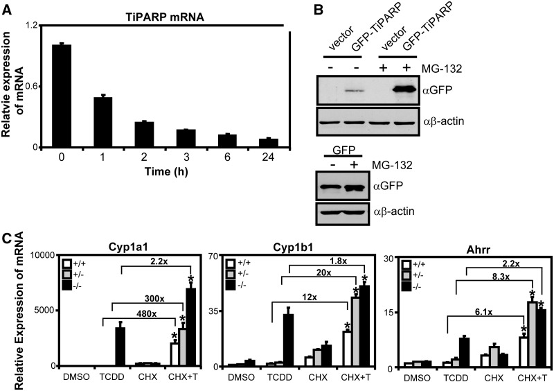
Figure 8.
TiPARP is a labile negative regulator of AHR. (A) TiPARP mRNA was rapidly degraded after actinomycin D treatment. T-47D cells were treated with 1 µg/ml actinomycin D at the times indicated and Tiparp mRNA expression levels determined by qPCR. (B) GFP-TiPARP overexpression was increased after proteasome inhibition. HuH-7 cells were transfected with pEGFP-TiPARP, pcDNA (vector) and pEGFP for 24 h and treated with 25 µM MG-132 for 6 h, and GFP-TiPARP protein levels were determined by western blot using anti-GFP antibody. The data were from a representative western blot from three independent experiments. (C) Ahr target gene induction in MEF lines pre-treated with 10 µg/ml CHX. MEF cell lines were pre-treated with 10 µg/ml CHX for 1 h, then treated with TCDD for 6 h and gene expression was determined. Data were normalized to wildtype DMSO. Fold changes between TCDD alone and CHX + TCDD (CHX + T) were provided for each gene. Gene expression results were shown as means ± S.E.M for three independent experiments and significance analysed by one-way ANOVA and Tukey’s multiple comparisons test. Gene expression levels significantly different (P < 0.05) than TCDD alone within each genotype were denoted with an asterisk.

