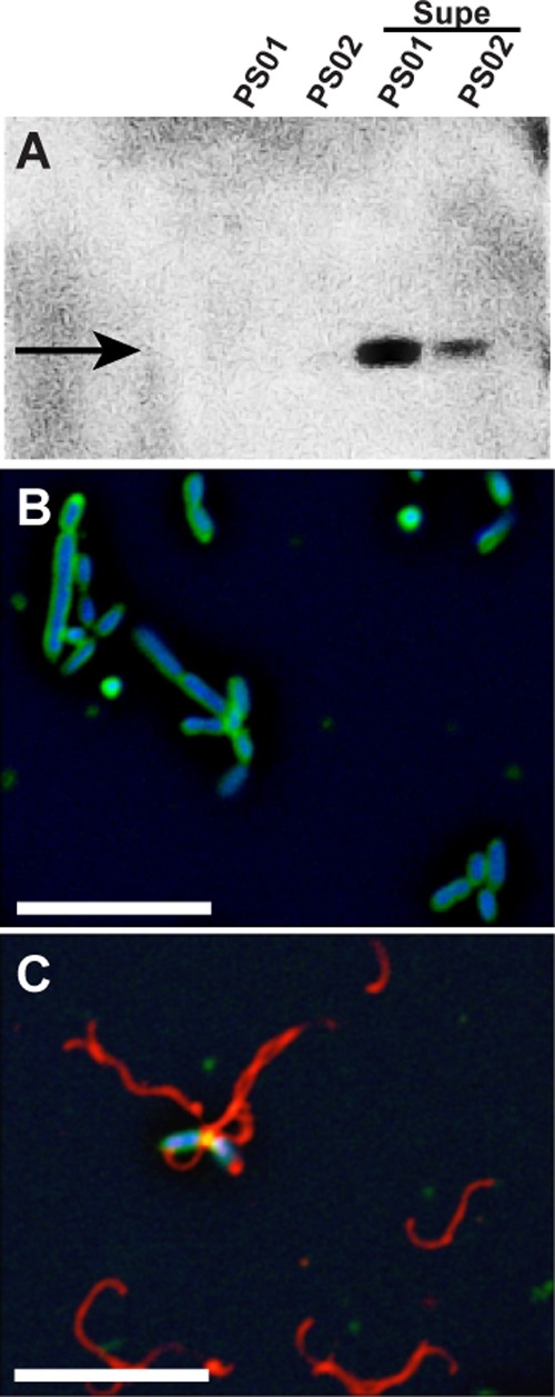
Fig 1.

Effect of RMI on flagella biosynthesis. (A) Immunoblot with anti-flagellin antiserum. Lanes, from left to right: PS01, untreated; PS02, untreated; PS01, treated with RMI supernatant; PS01, treated with RMI supernatant. The arrow indicates the major flagellin, FliC3. (B and C) Immunofluorescence microscopy of untreated PS01 cells (B) and RMI supernatant-treated PS01 cells (C). A cell-free RMI supernatant (Supe) was obtained from a coculture of PS01 and PS02 cells, and PS01 cells were treated by resuspending the cells in RMI supernatant and incubating for 90 min at 30°C. In panels B and C, flagella are false-colored red, membranes are green, and DNA is blue. Scale bar, 5 μm.
